Medical electronics
The calculated heart
Researchers at Karlsruher Institut für Technologie (KIT) are working on realistic computer models of the heart. They simulate physiological and pathological processes and develop models to individually assess the risk of cardiac arrhythmias and the effect of therapies.
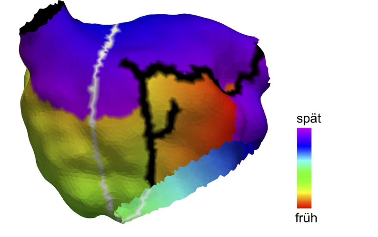
The risk of a patient developing atypical atrial flutter has not yet been reliably investigated. Researchers at the Karlsruher Institut für Technologie, the Medical Hospital IV of the Karlsruhe Municipal Hospital, the Medical Faculty of the University of Freiburg and the University Heart Centre Freiburg - Bad Krozingen have now developed a method to assess the risk of atypical atrial flutter. Using personalised computer models, they identify all the paths along which the atypical, circular electrical excitations can occur.
»Our models incorporate anatomical, electrophysiological and pharmacological criteria,« explains Dr. Axel Loewe, head of the cardiac modeling workgroup at the Institute of Biomedical Engineering at KIT. The effect of therapies such as catheter ablation or medications can also be individually assessed in advance.
The work demonstrates the advantages of mathematically simulated organs for medicine. »Computer models offer a perfectly controllable environment for experiments,« explains Loewe. »This allows individual changes to be simulated and their consequences for the overall system to be calculated.« The models complement classical methods such as cell and animal experiments and make it possible to test new therapies without risk for humans.
Personalized models reduce risk

Loewe already simulated the causes of atrial fibrillation with the computer in his dissertation. The KIT cardiac modelling group, which he heads, develops realistic models of the heart at all levels from the ion channel to cells and tissue to the complete organ. They can simulate how electrical excitation develops, spreads through the atria and the entire heart and – in the case of a healthy beating heart – extinguishes or – in the case of certain cardiac arrhythmias – sustains itself permanently.
In addition to simulating such basic physiological and pathological processes, the group is also working on personalized models to determine the risk of disease and the effect of treatments individually. In order to determine the personal anatomy of a patient, such as the size and shape of the atria, the researchers use imaging techniques such as magnetic resonance imaging. The group works closely with the Bioelectrical Signals working group at the Institute of Biomedical Engineering at the KIT, headed by Professor Olaf Dössel, on the inclusion of the electrical activity of the heart recorded by electrocardiogram (ECG).


